Clarius Mobile Health has introduced T-Mode Heart, an innovative AI-powered educational tool designed to help primary care physicians and non-cardiologists quickly learn and perform cardiac ultrasound (echocardiography). The solution overlays real-time anatomical labels, measurements, and guidance directly on the ultrasound image, significantly shortening the learning curve and improving diagnostic confidence for frontline clinicians.
Glimpse:
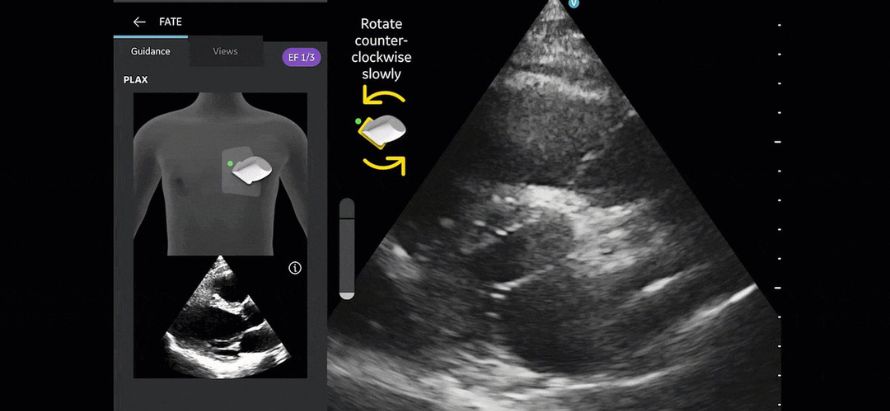
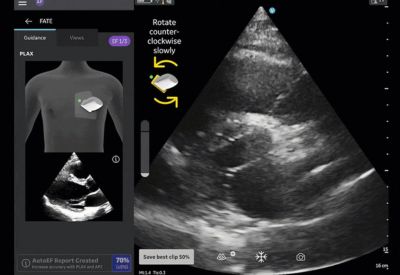
T-Mode Heart uses artificial intelligence to automatically recognize and label key cardiac structures (chambers, valves, pericardium, etc.) in real time during live scanning. It provides visual guidance, standard measurement tools, and educational overlays to assist users in acquiring high-quality cardiac views and interpreting basic findings. The tool is integrated into Clarius handheld ultrasound scanners and is aimed at primary care physicians, emergency medicine doctors, internal medicine specialists, and other non-cardiologists who increasingly need to perform bedside cardiac ultrasound for rapid assessment of heart function, fluid status, and structural abnormalities.
Clarius Mobile Health, a global leader in handheld ultrasound technology, has launched T-Mode Heart, a groundbreaking AI educational and guidance tool that enables primary care physicians and other non-cardiologists to confidently perform and interpret cardiac ultrasound examinations. Unveiled at a major medical imaging conference, the new feature addresses a critical gap in point-of-care ultrasound adoption: the steep learning curve associated with acquiring and interpreting high-quality cardiac views, which traditionally required extensive cardiology training.
T-Mode Heart works by applying advanced AI models to live ultrasound images in real time. As the clinician scans the patient’s heart, the system automatically recognizes and labels essential anatomical structures including the left ventricle, right ventricle, atria, valves, pericardium, and major vessels while providing visual guidance on probe positioning and image optimization. The tool also displays standard cardiac measurements (ejection fraction estimation, chamber dimensions, and pericardial effusion assessment) and offers educational overlays explaining normal versus abnormal findings, helping users build competence rapidly.
The solution is fully integrated into Clarius’s wireless handheld ultrasound scanners, requiring no additional hardware or complex setup. It supports multiple cardiac windows (parasternal long-axis, short-axis, apical four-chamber, and subcostal views) and is designed to work in both emergency and routine primary care settings. Early feedback from pilot users indicates that T-Mode Heart significantly reduces the time required to acquire diagnostic-quality cardiac images and improves diagnostic confidence among non-specialist physicians.
Clarius executives noted that the launch of T-Mode Heart is part of the company’s broader mission to democratize ultrasound by making it more accessible and usable for frontline clinicians. With the growing adoption of point-of-care ultrasound (POCUS) in primary care, emergency medicine, and rural health settings, tools that lower the barrier to performing and interpreting cardiac scans are becoming increasingly important for timely diagnosis of heart failure, valvular disease, pericardial effusion, and other common cardiac conditions.
The T-Mode Heart feature is now available as an optional add-on for Clarius cardiac and general-purpose handheld scanners. The company plans to expand the T-Mode series with additional educational AI tools for other anatomical regions in the near future.