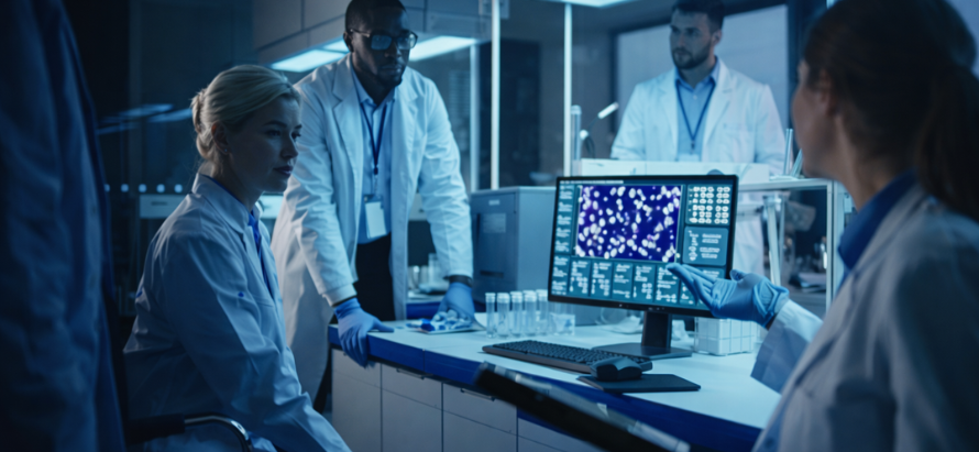
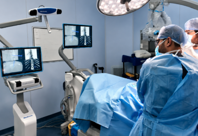
AI integration in radiology and pathology has reached a tipping point in India, with widespread adoption of deep learning models for faster, more accurate interpretation of X-rays, CTs, MRIs, mammograms, histopathology slides, and cytology smears. These tools are reducing diagnostic turnaround times, improving detection rates for high-burden conditions (TB, lung cancer, diabetic retinopathy, breast cancer), minimising inter-observer variability, and enabling radiologists and pathologists to focus on complex cases particularly in tier-2/3 cities and public-sector facilities facing severe specialist shortages.
Glimpse:
By early 2026, over 500 hospitals, diagnostic chains, and public health programmes across India are using AI solutions from Qure.ai, SigTuple, Niramai, DeepTek, Artelus, and others. Key applications include AI-assisted chest X-ray screening for TB and pneumonia, automated detection of diabetic retinopathy in tele-ophthalmology, breast cancer risk stratification via thermal/mammography AI, and digital pathology for faster slide analysis. Real-world studies show 30–60% reductions in reporting time, 10–25% improvement in early detection sensitivity, and significant workload relief for overburdened radiologists and pathologists.
The convergence of abundant medical imaging data, affordable cloud computing, and India-specific AI models has propelled AI from experimental pilots to mainstream deployment in radiology and pathology departments across the country. In 2026, AI is no longer a futuristic add on it is becoming essential infrastructure for handling India’s massive diagnostic workload amid a persistent shortage of specialists.
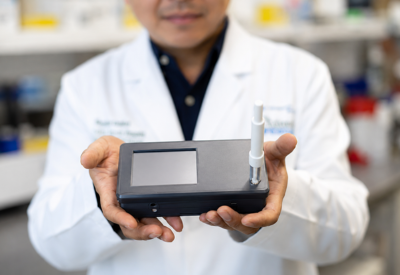
In radiology, AI tools now routinely analyse chest X-rays for tuberculosis, pneumonia, and lung nodules (Qure.ai, DeepTek), mammograms and breast thermal images for cancer (Niramai, Artelus), retinal scans for diabetic retinopathy (Remidio, Forus), and CT/MRI scans for stroke, trauma, and neurological emergencies. These systems function as “second readers” or triage assistants, flagging urgent cases for immediate human review while clearing normal or low-probability studies quickly. In high-volume public hospitals and district facilities, this has reduced average chest X-ray reporting time from hours to minutes, enabling faster initiation of TB treatment and reducing loss to follow-up.
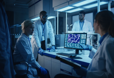
Digital pathology is seeing parallel transformation. AI-assisted slide analysis platforms (SigTuple, Morphle Labs, Abridge) automatically screen peripheral blood smears, Pap smears, histopathology slides, and frozen sections for abnormalities—identifying malaria parasites, malignant cells, grading tumours, and quantifying mitotic activity with high reproducibility. In cancer centres and large diagnostic chains, these tools have cut turnaround times for oncology biopsies from days to hours, improved consistency in grading, and allowed pathologists to focus on challenging cases rather than routine screening.
The impact is most pronounced in tier-2/3 cities and rural outreach programmes, where radiologist and pathologist shortages are acute. AI enables task-shifting: trained technicians or general physicians can perform initial scans or slide preparation, with AI providing preliminary reports that are later over-read by specialists via tele-radiology or tele-pathology. This model has proven especially effective in national programmes such as TB screening, diabetic retinopathy screening under NPCDCS, and cervical cancer screening under the National Programme for Prevention and Control of Cancer.
Real-world evidence is mounting. Multiple Indian studies and deployments have shown:
- 30–60% reduction in radiology reporting time
- 10–25% improvement in early detection sensitivity for TB, DR, and breast cancer
- Significant reduction in inter observer variability in pathology grading
- Lower rates of missed findings in high-volume, fatigue prone settings
- Cost savings from reduced repeat testing and unnecessary referrals
Challenges persist ensuring algorithmic fairness across diverse Indian skin tones, body habitus, and disease patterns; maintaining clinician trust through explainability and transparency; addressing data privacy under the DPDP Act; and building sustainable economics for public-sector adoption. Yet the trajectory is clear: AI-supported radiology and pathology are transitioning from nice-to-have enhancements to essential tools for managing India’s diagnostic workload efficiently and equitably.